The liver is one of the most remarkable organs in the human body. It is a masterful biochemical factory that performs hundreds of vital functions. This comprehensive guide explores the intricate anatomy and physiology of the liver, providing essential insights into its structure and functions.
Understanding Liver Anatomy
The liver, the largest internal organ in the human body, occupies a strategic position in the upper right portion of the abdominal cavity, just beneath the diaphragm. This reddish-brown organ weighs approximately 1.5 kilograms (3.3 pounds) in adults and extends across the entire abdominal cavity from right to left.
Structural Organization
The liver’s architecture follows a complex yet organized pattern. The organ consists of two primary lobes – a larger right lobe and a smaller left lobe. These main lobes further divide into eight functional segments, each receiving blood supply and biliary drainage. This segmental organization allows surgeons to remove portions of the liver while maintaining the organ’s essential functions.
Within each segment, thousands of hexagonal-shaped functional units called liver lobules exist. These lobules contain specialized liver cells (hepatocytes) arranged in plates that radiate from a central vein, similar to the spokes in a wheel. This unique arrangement optimizes the liver’s ability to process blood and produce bile.
Blood Supply System
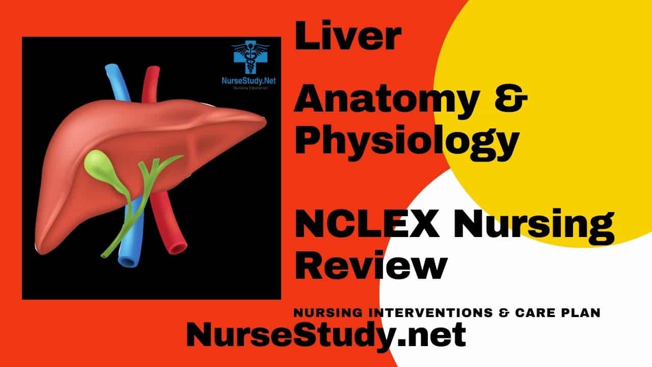
The liver’s blood supply system stands out as unique among all organs, receiving blood from two distinct sources:
- The hepatic portal vein delivers nutrient-rich blood from the digestive tract and spleen, accounting for about 75% of the liver’s blood supply
- The hepatic artery provides oxygen-rich blood, making up the remaining 25%
This dual blood supply ensures that the liver receives the oxygen and nutrients necessary to perform its numerous functions while also processing materials absorbed from the digestive tract.
Essential Liver Physiology
The liver’s physiological functions span a remarkable range of activities, making it one of the body’s most versatile organs. Here are its key functions:
Metabolism and Nutrient Processing
The liver plays a central role in:
- Carbohydrate metabolism: Converting excess glucose to glycogen for storage and maintaining blood glucose levels
- Protein synthesis: Producing essential proteins for blood plasma, including clotting factors
- Lipid processing: Manufacturing cholesterol and specialized proteins for fat transport throughout the body
- Vitamin storage: Maintaining reserves of vitamins A, D, E, K, and B12
Detoxification and Waste Management
The liver serves as the body’s primary detoxification center by:
- Converting toxic ammonia to urea for safe excretion
- Processing drugs and medications into less harmful compounds
- Filtering bacteria and pathogens from the blood
- Breaking down old or damaged red blood cells
Bile Production and Secretion
The liver produces approximately 500-1000 mL of bile daily, which serves two essential purposes:
- Facilitating fat digestion in the small intestine
- Enabling the excretion of waste products, including bilirubin from broken-down red blood cells
Immune Function
The liver contributes significantly to the body’s immune defense by:
- Producing immune factors and proteins
- Removing pathogens from the bloodstream
- Housing specialized immune cells called Kupffer cells
Clinical Significance
Understanding liver anatomy and physiology is crucial for:
- Diagnosing and treating liver diseases
- Planning surgical procedures
- Developing new therapeutic approaches
- Monitoring liver function in patients with chronic conditions
The liver’s remarkable regenerative capacity allows it to function even when up to 75% of its tissue is damaged or removed, highlighting its resilience and importance to survival.
References:
- Trefts E, Gannon M, Wasserman DH. The liver. Curr Biol. 2019;29(21):R1019-R1024. doi:10.1016/j.cub.2019.09.017
- Kietzmann T. Metabolic zonation of the liver: The oxygen gradient revisited. Redox Biol. 2017;11:622-630. doi:10.1016/j.redox.2017.01.012
- Matsumoto T, Kawakami M. The unit-concept of hepatic parenchyma–a re-examination based on angioarchitectural studies. Acta Pathol Jpn. 2018;32 Suppl 2:285-314.
- Boyer TD, Wright TL, Manns MP. Zakim and Boyer’s Hepatology: A Textbook of Liver Disease, 6th ed. Philadelphia: Saunders; 2022.