Cellulitis is a common and potentially serious bacterial skin infection affecting the dermis and deeper subcutaneous tissue layers. Most cases are caused by Staphylococcus aureus (including methicillin-resistant strains, MRSA) or beta-hemolytic streptococci such as Streptococcus pyogenes, which invade through breaks in the skin barrier—cuts, abrasions, insect bites, surgical wounds, or chronic ulcers.
Without prompt recognition and treatment, cellulitis can rapidly progress from localized redness and swelling to life-threatening systemic infection, including sepsis, necrotizing fasciitis, and bacteremia.
For nursing students preparing for the NCLEX and nurses working in emergency departments, medical-surgical units, or community health settings, understanding the nursing diagnosis for cellulitis is fundamental to creating evidence-based care plans, preventing complications, and promoting patient recovery. This guide provides comprehensive assessment strategies, appropriate NANDA nursing diagnoses, detailed interventions with rationales, and five complete nursing care plan examples to support safe, effective clinical practice.
Definition and Overview
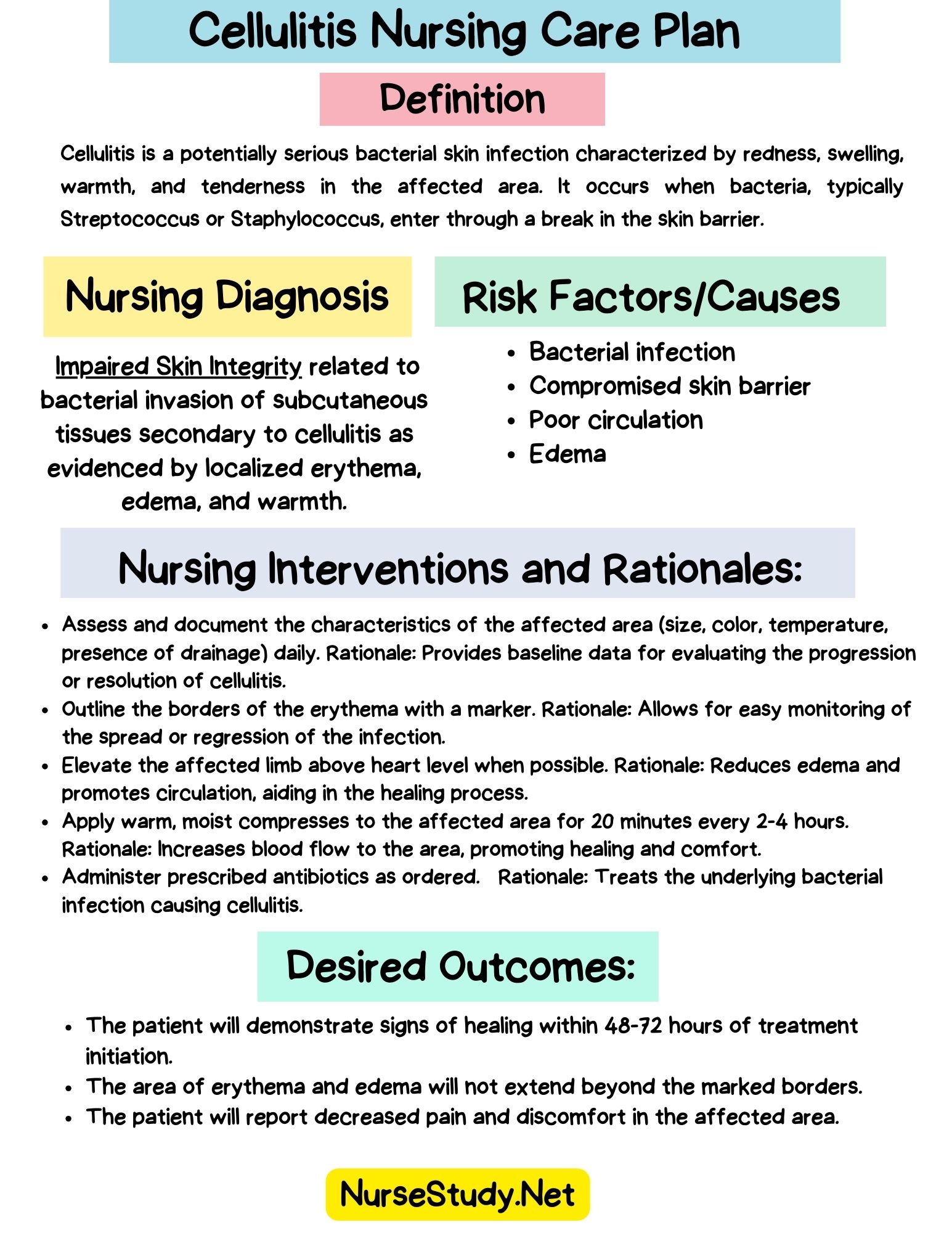
Cellulitis is an acute, spreading bacterial infection of the skin and subcutaneous tissues characterized by localized warmth, erythema, edema, and pain. Unlike superficial infections such as impetigo or folliculitis, cellulitis penetrates deeper into dermal and subcutaneous layers, causing significant inflammation and tissue damage.
The condition most commonly affects the lower extremities, particularly the legs and feet, but can occur anywhere on the body. Facial cellulitis (especially periorbital) requires urgent evaluation due to the risk of intracranial complications.
In clinical practice, nurses frequently encounter cellulitis in patients with diabetes, peripheral vascular disease, lymphedema, obesity, or immunocompromising conditions—all factors that increase susceptibility and complicate treatment. Early identification and aggressive antibiotic therapy are essential to prevent progression to severe complications including abscess formation, osteomyelitis, endocarditis, and septic shock.
Pathophysiology
Cellulitis develops when bacteria breach the protective skin barrier and invade deeper tissue layers, triggering a robust inflammatory cascade.
Mechanism of Infection
Bacterial Entry: Pathogens enter through disruptions in skin integrity—lacerations, puncture wounds, surgical incisions, ulcers, fissures between toes, insect bites, or areas of maceration from moisture or friction. In patients with chronic edema or lymphedema, the skin becomes fragile and more susceptible to minor trauma, which allows bacterial entry.
Inflammatory Response: Once bacteria invade the dermis and subcutaneous tissue, the immune system mounts an inflammatory response. Neutrophils and macrophages migrate to the site, releasing cytokines and inflammatory mediators. This cascade causes vasodilation, increased vascular permeability, and fluid extravasation into surrounding tissues.
Clinical Manifestations: Vasodilation produces the characteristic erythema (redness) and warmth. Plasma leakage into interstitial spaces causes edema and swelling. Bacterial toxins and inflammatory mediators stimulate pain receptors, resulting in tenderness and discomfort. The affected area often has poorly defined borders that advance as infection spreads along tissue planes.
Systemic Complications
If untreated or inadequately treated, cellulitis can spread through several pathways:
- Lymphatic spread: Bacteria enter lymphatic channels, causing lymphangitis (visible red streaks tracking proximally) and regional lymphadenopathy. Continued spread can lead to systemic infection.
- Hematogenous spread: Bacteria invade blood vessels, resulting in bacteremia and potential seeding of distant organs (endocarditis, osteomyelitis, septic arthritis).
- Deep tissue extension: Infection can track along fascial planes, leading to necrotizing fasciitis—a surgical emergency with high mortality requiring immediate debridement.
- Abscess formation: Localized collections of purulent material may develop, requiring incision and drainage in addition to antibiotics.
Patients with diabetes face a particularly high risk due to hyperglycemia impairing neutrophil function, microvascular disease reducing tissue perfusion and antibiotic delivery, and peripheral neuropathy delaying recognition of early infection signs.
Causes and Related Factors
Understanding the etiology and risk factors for cellulitis guides nursing assessment priorities and patient education.
Common Bacterial Pathogens
- Beta-hemolytic streptococci (Streptococcus pyogenes, Group A Strep): Most common cause of non-purulent cellulitis
- Staphylococcus aureus: Including methicillin-resistant strains (MRSA), especially in purulent cellulitis or with abscess formation
- Less common organisms: Streptococcus pneumoniae, Haemophilus influenzae (facial cellulitis in children), Pseudomonas aeruginosa (immunocompromised patients, aquatic exposures), Vibrio vulnificus (saltwater exposure with open wounds)
Predisposing Factors (NANDA “Related To” Factors)
Skin barrier disruption:
- Traumatic injuries (cuts, abrasions, puncture wounds, bites)
- Surgical incisions or invasive procedures
- Chronic wounds (venous stasis ulcers, pressure injuries, diabetic foot ulcers)
- Dermatologic conditions (eczema, psoriasis, athlete’s foot, tinea pedis)
- Intravenous drug use (injection sites)
Vascular and lymphatic compromise:
- Chronic venous insufficiency
- Peripheral arterial disease
- Lymphedema (post-mastectomy, chronic leg edema, filariasis)
- Previous episodes of cellulitis (damages lymphatic drainage)
Metabolic and immune factors:
- Diabetes mellitus (especially poor glycemic control with HbA1c >7.5%)
- Obesity
- Immunosuppression (chemotherapy, corticosteroids, HIV/AIDS, organ transplant)
- Chronic kidney disease
- Cirrhosis or liver disease
- Malnutrition
Environmental and behavioral factors:
- Poor hygiene practices
- Walking barefoot
- Occupational exposures (agricultural work, aquatic environments)
- History of recurrent cellulitis
Research demonstrates that patients with diabetes have up to 1.4 times higher risk of cellulitis, with risk increasing 12% for every 1% elevation in HbA1c. This underscores the critical importance of glucose management in infection prevention.
Signs and Symptoms
Thorough assessment of both subjective reports and objective findings is essential for accurate nursing diagnosis and monitoring treatment response.
Subjective Data (What Patients Report)
- Pain, tenderness, or aching in the affected area
- Sensation of warmth or “heat” in the skin
- Progressive worsening over hours to days
- Recent skin trauma, bite, or injury
- Fever, chills, or generalized malaise
- History of similar episodes
Objective Data (What Nurses Observe and Measure)
Local signs:
- Erythema (redness) with poorly defined, irregular borders
- Edema and swelling of the affected tissue
- Warmth to touch (increased skin temperature)
- Tenderness on palpation
- Tense, shiny skin appearance
- Possible purulent drainage if an abscess is present
- Skin breakdown, ulceration, or open wound
- Red streaks extending proximally (lymphangitis)
Systemic signs:
- Fever (often >38°C/100.4°F in moderate to severe cases)
- Tachycardia
- Regional lymphadenopathy (enlarged, tender lymph nodes)
- Malaise and fatigue
- Hypotension (if progressing to sepsis)
Diagnostic Findings
Laboratory tests:
- Elevated white blood cell count (WBC): Leukocytosis with left shift indicates acute bacterial infection
- Elevated inflammatory markers: C-reactive protein (CRP) and erythrocyte sedimentation rate (ESR)
- Blood cultures: Obtained if systemic symptoms present; positive in bacteremia
- Wound or aspirate culture: May be performed in purulent cellulitis, but routine culture of non-purulent cellulitis has low yield
Imaging studies:
- Ultrasound: May be ordered to rule out abscess or deep vein thrombosis
- CT or MRI: Reserved for severe cases or when necrotizing fasciitis suspected
Red-Flag Assessment Findings
Nurses must recognize warning signs requiring urgent physician notification or escalation of care:
- Rapidly spreading erythema (progression beyond marked borders within hours)
- Blistering, skin necrosis, or crepitus (gas in tissues)
- Severe pain out of proportion to physical findings
- Signs of sepsis (hypotension, altered mental status, tachycardia, tachypnea)
- Periorbital or facial cellulitis (risk of cavernous sinus thrombosis)
- Lack of improvement after 48-72 hours of appropriate antibiotic therapy
- Extreme tenderness, fever >39°C, or systemic toxicity
Expected Outcomes and Goals
Effective nursing care plans include measurable, patient-centered outcomes consistent with Nursing Outcomes Classification (NOC) language.
Short-Term Goals (Within 48-72 Hours)
- Patient will verbalize pain reduction to ≤3/10 on the numeric rating scale
- Erythema will demonstrate no further advancement beyond marked borders
- Patient will remain afebrile (temperature <37.8°C/100°F)
- Patient will verbalize understanding of the antibiotic regimen and complete the first 48 hours of therapy
- Edema will decrease, as evidenced by reduced circumference measurements
Long-Term Goals (By Discharge/End of Treatment Course)
- Skin integrity will be restored with no open wounds, drainage, or erythema
- Patient will demonstrate proper wound care technique and hand hygiene
- White blood cell count will return to normal limits (4,000-11,000/μL)
- Patient will identify personal risk factors and prevention strategies
- Patient will verbalize signs/symptoms requiring immediate medical attention
- Patient will complete full prescribed antibiotic course (typically 5-14 days)
Nursing Assessment Priorities
Systematic assessment guides accurate nursing diagnosis and monitors treatment effectiveness.
History Taking
Present illness:
- Onset and duration of symptoms
- Progression pattern (how quickly spreading)
- Location and characteristics of initial skin injury
- Previous antibiotic use or allergies
- Recent travel, exposures, or insect/animal bites
Medical history:
- Diabetes and current glucose control
- Peripheral vascular disease or venous insufficiency
- Previous episodes of cellulitis or skin infections
- Immunocompromising conditions
- Lymphedema or lymphatic surgery
- Chronic wounds or pressure injuries
- Dermatologic conditions
Medication review:
- Immunosuppressive medications (corticosteroids, chemotherapy)
- Anticoagulants (may affect treatment decisions)
- Current antibiotic therapy
Physical Examination
Integumentary assessment:
- Mark the borders of erythema with pen/marker and date/time to monitor progression or regression
- Measure the circumference of the affected limb at a consistent landmark to track edema
- Assess skin temperature using the back of the hand, comparing the affected to the unaffected side
- Evaluate skin integrity, noting any breaks, ulcers, or drainage
- Look for entry point (tinea pedis between toes, puncture wound, insect bite)
- Check for blistering, purpura, or necrotic tissue (concerning signs)
Vascular assessment:
- Palpate peripheral pulses (dorsalis pedis, posterior tibial, or appropriate to location)
- Assess capillary refill time (normal <3 seconds)
- Evaluate the color and temperature of digits
- Note any cyanosis or mottling
Lymphatic assessment:
- Palpate regional lymph nodes for enlargement and tenderness
- Observe for red streaking (lymphangitis)
Systemic assessment:
- Vital signs every 4 hours minimum; monitor for fever, tachycardia, hypotension
- Mental status (confusion may indicate sepsis)
- Urine output (decreased output may signal sepsis or dehydration)
Pain Assessment
Use a standardized pain scale (0-10 numeric rating scale) and assess:
- Intensity, quality, location
- Aggravating and relieving factors
- Impact on function and mobility
- Response to analgesic interventions
Psychosocial Assessment
- Patient’s understanding of diagnosis and treatment plan
- Anxiety or fear related to infection or hospitalization
- Barriers to medication adherence (cost, transportation, health literacy)
- Home environment and self-care ability
- Support system and resources
Nursing Interventions and Rationales
These core interventions apply broadly to cellulitis management and support multiple nursing diagnoses. Specific interventions tailored to individual nursing diagnoses appear in the care plan examples below.
Infection Management
Administer prescribed antibiotic therapy on schedule.
Rationale: Maintaining therapeutic antibiotic levels is essential to eradicate bacteria and prevent progression. Oral antibiotics (cephalexin, dicloxacillin, clindamycin) are appropriate for mild to moderate cellulitis. IV antibiotics (vancomycin, cefazolin, piperacillin-tazobactam) are indicated for severe infection, systemic symptoms, facial cellulitis, failed outpatient treatment, or MRSA coverage.
Monitor for antibiotic effectiveness and adverse reactions.
Rationale: Improvement should be evident within 48-72 hours (decreased pain, reduced fever, no advancement of erythema). Lack of improvement warrants culture, imaging, or antibiotic adjustment. Common adverse reactions include GI upset, rash, or Clostridioides difficile infection.
Perform meticulous hand hygiene before and after patient contact and wound care.
Rationale: Hand hygiene is the single most effective measure to prevent healthcare-associated infections and cross-contamination between patients.
Use appropriate isolation precautions.
Rationale: Contact precautions may be indicated for draining wounds or suspected MRSA colonization to prevent transmission to other patients or healthcare workers.
Wound and Skin Care
Inspect the affected area daily and document characteristics: size, color, temperature, drainage, and borders.
Rationale: Serial assessments track progression or resolution and identify complications. Dating and marking borders provides objective evidence of treatment response.
Keep the affected area clean and dry; cleanse with mild soap and water.
Rationale: Gentle cleansing removes debris and exudate without causing additional trauma. Harsh antiseptics may damage healing tissue.
Apply sterile dressings to open wounds or areas with drainage.
Rationale: Dressings protect wounds from contamination, absorb drainage, and maintain moist healing environment. Change dressings per facility protocol or when saturated.
Avoid restrictive clothing, tight bandages, or constrictive devices on affected limb.
Rationale: Constriction impairs circulation and lymphatic drainage, worsening edema and potentially compromising tissue perfusion.
Edema and Circulation Management
Elevate affected extremity above heart level when possible.
Rationale: Elevation promotes venous return and lymphatic drainage, reducing edema and tissue pressure. This decreases pain and improves antibiotic penetration to infected tissues.
Encourage gentle range-of-motion exercises when appropriate.
Rationale: Movement promotes circulation without causing injury. Muscle contraction assists venous return. Avoid aggressive exercise during the acute phase.
Assess peripheral perfusion: pulses, capillary refill, color, temperature, sensation.
Rationale: Severe edema can compress vessels and compromise perfusion. Early detection of perfusion changes prevents ischemic injury.
Pain and Comfort Management
Administer prescribed analgesics (acetaminophen, NSAIDs, opioids for severe pain) and evaluate effectiveness.
Rationale: Pain control improves patient comfort, promotes rest and healing, and facilitates participation in care. NSAIDs provide both analgesic and anti-inflammatory effects.
Apply cool or warm compresses as ordered and tolerated.
Rationale: Cool compresses may reduce inflammation and pain in early stages; warm compresses may improve circulation and comfort in later stages. Follow provider orders, as heat is sometimes contraindicated in acute inflammation.
Promote rest and limit ambulation during the acute phase.
Rationale: Rest reduces metabolic demands, decreases tissue edema from dependent positioning, and conserves energy for immune response.
Monitoring for Complications
Monitor vital signs and assess for signs of systemic infection or sepsis.
Rationale: Fever, tachycardia, hypotension, altered mental status, or decreased urine output may indicate bacteremia or sepsis requiring immediate intervention and possible ICU-level care.
Monitor laboratory values: WBC, CRP, blood cultures, and renal function.
Rationale: Trending inflammatory markers guide treatment effectiveness. Renal function monitoring is important because some antibiotics are nephrotoxic and require dose adjustment.
Report red-flag findings immediately: rapidly spreading erythema, skin necrosis, severe pain, systemic toxicity.
Rationale: These signs may indicate necrotizing fasciitis, abscess, or sepsis—emergencies requiring urgent surgical or intensive care.
Patient Education
Teach the importance of completing the full antibiotic course, even if symptoms improve.
Rationale: Premature discontinuation leads to treatment failure, recurrence, and antibiotic resistance. Typical courses range from 5 to 14 days, depending on severity.
Educate on proper wound care, dressing changes, and hand hygiene.
Rationale: Correct technique prevents reinfection and promotes healing. Return demonstration ensures competency.
Discuss prevention strategies: skin protection, prompt wound care, and managing chronic conditions.
Rationale: Recurrence rates are significant (8-20% within 6 months). Prevention education addresses modifiable risk factors.
Instruct on signs requiring immediate medical attention: spreading redness, fever, increased pain, and red streaks.
Rationale: Early recognition and treatment of worsening infection prevent serious complications.
Nursing Care Plans for Cellulitis
Nursing Care Plan #1: Impaired Skin Integrity
Nursing Diagnosis Statement:
Impaired Skin Integrity related to bacterial invasion and inflammatory destruction of dermal and subcutaneous tissue secondary to cellulitis, as evidenced by erythema measuring 8 cm × 6 cm on right lower leg, edema, warmth to touch, purulent drainage from 2 cm break in skin, and patient report of tenderness rated 6/10.
Related Factors:
- Bacterial colonization and tissue invasion
- Inflammatory process causing tissue damage
- Compromised circulation or lymphatic drainage
- Presence of chronic wound or skin breakdown
- Diabetes with impaired healing
As Evidenced By:
- Erythema with irregular, poorly defined borders
- Localized edema and swelling
- Break in skin integrity (puncture wound, ulcer, or fissure)
- Purulent or serous drainage
- Warmth and tenderness on palpation
- Patient verbalizes pain at site
Expected Outcomes:
- Skin erythema will decrease by 50% within 72 hours, as measured by border markings
- Skin integrity will be restored with complete re-epithelialization within 10-14 days
- Patient will demonstrate proper wound cleansing and dressing technique before discharge
- Patient will verbalize three strategies to prevent skin breakdown and infection recurrence
Nursing Interventions and Rationales:
- Assess and document wound characteristics daily: size, depth, borders, drainage type and amount, and surrounding tissue condition. Mark erythema borders with dated/timed pen markings.
- Rationale: Serial documentation provides objective evidence of healing progression or deterioration. Marked borders allow nurses and providers to quickly identify spread of infection or response to treatment. Purulent drainage may indicate abscess requiring drainage.
- Cleanse affected area gently with mild soap and water or normal saline; pat dry thoroughly.
- Rationale: Gentle cleansing removes bacteria, exudate, and debris without traumatizing fragile healing tissue. Thorough drying prevents maceration. Harsh antiseptics (hydrogen peroxide, povidone-iodine) may damage new epithelial cells.
- Apply prescribed topical antimicrobial agents (mupirocin, silver sulfadiazine) and sterile dressings per protocol.
- Rationale: Topical antimicrobials may be ordered for localized wound infection. Sterile dressings protect the wound from external contamination, absorb exudate, and maintain moisture balance conducive to healing.
- Elevate affected extremity above heart level continuously during acute phase, then intermittently throughout day.
- Rationale: Elevation uses gravity to enhance venous return and lymphatic drainage, reducing interstitial edema. Decreased tissue pressure improves perfusion, oxygen delivery, and antibiotic penetration—all essential for healing.
- Instruct patient to avoid scratching, picking, or touching lesions; keep fingernails short and clean.
- Rationale: Scratching introduces additional bacteria, causes mechanical trauma, and creates new portals for infection. Short, clean nails reduce microbial load and tissue damage if scratching occurs.
- Teach patient proper skin care: daily inspection, moisturizing dry skin, treating athlete’s foot, prompt attention to cuts/abrasions.
- Rationale: Preventive skin care maintains barrier function. Dry, cracked skin and fungal infections (tinea pedis) are common entry points for cellulitis-causing bacteria. Early treatment of minor injuries prevents progression to cellulitis.
Nursing Care Plan #2: Acute Pain
Nursing Diagnosis Statement:
Acute Pain related to inflammatory process, tissue swelling, and bacterial toxins secondary to cellulitis, as evidenced by patient report of throbbing pain rated 7/10 on numeric scale, facial grimacing, guarding of affected right lower leg, reluctance to ambulate, and elevated heart rate of 98 bpm at rest.
Related Factors:
- Inflammation and vasodilation cause tissue pressure
- Edema compresses nerve endings
- Bacterial toxins irritate pain receptors
- Tissue ischemia from compromised perfusion
As Evidenced By:
- Verbal report of pain with numeric rating (typically 4-8/10)
- Facial expressions of pain (grimacing, frowning, tense appearance)
- Guarding behavior (protecting or favoring the affected area)
- Restlessness or inability to find a comfortable position
- Autonomic responses (tachycardia, elevated blood pressure)
- Decreased functional ability (reluctance to move or bear weight)
Expected Outcomes:
- Patient will report pain decreased to ≤3/10 within 48-72 hours of treatment initiation
- Patient will demonstrate relaxed body posture and facial expression
- Patient will sleep without pain-related interruptions
- Patient will participate in activities of daily living and ambulation with minimal discomfort
- Patient will verbalize effective pain relief strategies, including both pharmacologic and non-pharmacologic methods
Nursing Interventions and Rationales:
- Assess pain systematically every 4 hours and PRN using numeric rating scale (0-10). Document intensity, quality, location, duration, aggravating and relieving factors.
- Rationale: Standardized pain assessment provides baseline data and tracks treatment response. Pain intensity and quality help differentiate cellulitis pain from other causes (DVT, compartment syndrome, ischemia). Increased pain may signal complications.
- Administer prescribed analgesics on schedule: acetaminophen for mild pain; NSAIDs (ibuprofen, naproxen) for moderate pain with inflammation; opioids for severe pain. Evaluate effectiveness 30-60 minutes post-administration.
- Rationale: Scheduled dosing maintains consistent pain control better than PRN dosing. NSAIDs address both pain and inflammation. Acetaminophen also reduces fever, improving overall comfort. Evaluation ensures adequate relief and guides dose adjustments. Use lowest effective dose to minimize adverse effects.
- Elevate affected limb on pillows above heart level; maintain comfortable, supported position.
- Rationale: Elevation reduces hydrostatic pressure in capillaries, decreasing edema and tissue pressure. Reduced swelling decompresses nerve endings, providing pain relief. Proper positioning prevents additional discomfort from awkward limb angles.
- Apply warm compresses to affected area for 15-20 minutes three to four times daily if ordered (avoid if contraindicated by acute inflammation).
- Rationale: Warmth increases local blood flow, delivering oxygen and antibiotics to tissues while removing inflammatory mediators and metabolic waste. Improved circulation may reduce pain. Use cautiously—excessive heat can worsen inflammation in hyperacute phase. Verify orders and patient tolerance.
- Teach non-pharmacologic pain management techniques: deep breathing exercises, guided imagery, distraction (music, reading), progressive muscle relaxation.
- Rationale: Non-pharmacologic strategies complement medications and give patients active coping skills. Relaxation reduces muscle tension and anxiety, which amplify pain perception. Distraction shifts attention away from pain signals.
- Encourage adequate rest periods and minimize unnecessary movement during acute phase; assist with activities requiring ambulation on affected limb.
- Rationale: Rest reduces mechanical stress on inflamed tissues and conserves energy for healing. Dependent positioning from prolonged standing or ambulation increases edema and pain. Assistance prevents falls and injury while accommodating mobility limitations from pain.
Nursing Care Plan #3: Risk for Infection (Systemic Spread)
Nursing Diagnosis Statement:
Risk for Infection (systemic spread, sepsis) related to presence of virulent bacteria, compromised skin integrity, and potential for inadequate immune response in patient with diabetes mellitus type 2 and HbA1c of 9.2%.
Risk Factors:
- Existing localized infection (cellulitis)
- Diabetes with poor glycemic control (impaired neutrophil function)
- Obesity (adipose tissue relatively hypoperfused)
- Open wound providing portal for bacterial entry
- Delayed treatment initiation
- Potential antibiotic resistance (MRSA)
Expected Outcomes:
- Patient will remain free of signs of systemic infection (bacteremia, sepsis) throughout treatment course
- Patient will maintain temperature within normal limits (36.5-37.5°C/97.7-99.5°F)
- White blood cell count will trend downward toward normal range (4,000-11,000/μL) within 48-72 hours
- Blood cultures, if obtained, will be negative for bacterial growth
- Patient will complete prescribed antibiotic regimen as evidenced by self-report and pharmacy records
- Patient will verbalize understanding of infection prevention strategies
Nursing Interventions and Rationales:
- Perform strict hand hygiene with soap and water or alcohol-based sanitizer before and after all patient contact, wound care, or contact with potentially contaminated surfaces.
- Rationale: Hand hygiene is the single most critical intervention to prevent healthcare-associated infections and cross-contamination. Cellulitis pathogens can be transmitted via healthcare workers’ hands, leading to patient-to-patient spread or introduction of additional pathogens.
- Administer prescribed IV or oral antibiotic therapy precisely on schedule. Monitor for therapeutic response and adverse reactions (rash, diarrhea, nausea, nephrotoxicity).
- Rationale: Timely administration maintains therapeutic blood levels necessary to eradicate bacteria and prevent resistance development. Outpatient oral options include cephalexin 500 mg q6h, dicloxacillin 500 mg q6h, or clindamycin 300-450 mg q6-8h. Hospitalized patients or those with systemic symptoms require IV therapy (vancomycin for MRSA coverage, cefazolin, piperacillin-tazobactam). Monitoring identifies treatment failure or complications.
- Monitor vital signs every 4 hours minimum; immediately report fever >38.3°C (101°F), tachycardia >100 bpm, hypotension, tachypnea, or altered mental status.
- Rationale: These findings may indicate progression to bacteremia or sepsis—life-threatening conditions requiring immediate intervention. Early recognition and aggressive treatment (IV fluids, broad-spectrum antibiotics, possible ICU transfer) reduce mortality. Sepsis criteria include suspected infection plus two or more of: temperature >38°C or <36°C, heart rate >90 bpm, respiratory rate >20/min, WBC >12,000 or <4,000/μL.
- Monitor and trend laboratory values: daily CBC with differential, C-reactive protein (CRP), blood cultures if febrile or systemically ill.
- Rationale: Elevated and rising WBC with left shift (increased neutrophils and bands) indicates acute bacterial infection. Improving WBC suggests treatment effectiveness. CRP elevation confirms inflammation and trends with disease activity. Positive blood cultures identify causative organism and guide antibiotic selection based on susceptibilities.
- Educate patient on critical importance of completing entire antibiotic course even after symptoms resolve, typically 5-14 days depending on severity.
- Rationale: Premature antibiotic discontinuation allows surviving bacteria to proliferate, leading to relapse and promoting antibiotic resistance. Symptoms improve before bacteria are fully eradicated. Patient understanding and commitment to adherence are essential for cure.
- Teach patient to monitor for and immediately report warning signs: spreading redness, red streaks, worsening pain, fever, chills, confusion, dizziness.
- Rationale: These symptoms may indicate treatment failure, resistant organisms, or progression to systemic infection requiring urgent medical reevaluation. Early intervention prevents complications. Patients should understand when to call provider versus seeking emergency care.
Nursing Care Plan #4: Ineffective Peripheral Tissue Perfusion
Nursing Diagnosis Statement:
Ineffective Peripheral Tissue Perfusion related to inflammatory edema compressing blood vessels, pre-existing peripheral vascular disease, and diabetes-related microvascular damage, as evidenced by right lower leg edema measuring 4 cm greater circumference than left leg, capillary refill 4 seconds in right foot, diminished dorsalis pedis pulse, cool skin temperature distally, and patient report of numbness in toes.
Related Factors:
- Severe edema causing external compression of vessels
- Inflammation and endothelial dysfunction
- Pre-existing vascular insufficiency (arterial or venous disease)
- Diabetes with microvascular complications
- Immobility and limb dependency
- Potential deep vein thrombosis (DVT)
As Evidenced By:
- Edema and increased limb circumference
- Delayed capillary refill (>3 seconds)
- Diminished or absent peripheral pulses
- Cool skin temperature in distal extremity
- Pallor or cyanosis
- Numbness, tingling, or altered sensation
- Pain with passive muscle stretching
Expected Outcomes:
- Peripheral pulses will be palpable and equal bilaterally within 72 hours
- Capillary refill will improve to <3 seconds in affected extremity
- Edema will decrease by 50% as measured by daily circumference measurements
- Skin temperature and color will normalize in distal extremity
- Patient will verbalize decreased numbness or paresthesia
- Patient will demonstrate proper limb elevation technique
Nursing Interventions and Rationales:
- Assess peripheral perfusion every 4-8 hours: palpate pulses (dorsalis pedis, posterior tibial, popliteal, femoral as appropriate), assess capillary refill, evaluate skin color, temperature, and sensation. Compare affected to unaffected extremity.
- Rationale: Systematic vascular assessment identifies perfusion deficits and trends. Absent or diminished pulses, prolonged capillary refill, coolness, pallor, or cyanosis indicate arterial insufficiency. Severe edema can compress vessels, creating compartment-like syndrome. DVT must be ruled out (check for calf pain, Homans’ sign, asymmetric swelling). Early detection prevents ischemic injury.
- Measure and document circumference of affected limb at consistent anatomical landmark (e.g., 10 cm below tibial tuberosity) daily.
- Rationale: Objective measurements track edema progression or resolution better than subjective assessment. Decreased measurements indicate treatment effectiveness. Increasing measurements suggest worsening infection or inadequate treatment.
- Elevate affected extremity above heart level for majority of day; use pillows to support entire leg and prevent knee flexion contracture.
- Rationale: Elevation promotes venous and lymphatic drainage via gravity, reducing interstitial fluid accumulation. Decreased edema relieves vessel compression, improving blood flow. Proper support prevents pressure points and maintains neutral joint position.
- Encourage ankle pumps, dorsiflexion/plantarflexion exercises, and gentle ambulation as tolerated when acute inflammation subsides.
- Rationale: Muscle contraction acts as a pump, propelling venous blood proximally and reducing venous stasis. Gentle movement prevents DVT, maintains joint mobility, and improves circulation. Avoid vigorous exercise during acute inflammation.
- Avoid constrictive devices: tight bandages, elastic stockings, jewelry, or restrictive clothing on affected limb.
- Rationale: Constriction impedes arterial inflow and venous return, worsening edema and ischemia. Pressure from tight dressings can cause pressure injury to fragile, edematous skin.
- Monitor for compartment syndrome: severe pain disproportionate to findings, pain with passive stretch, paresthesia, paralysis, pulselessness, pallor. Report immediately if suspected.
- Rationale: Compartment syndrome is a surgical emergency caused by elevated pressure within fascial compartments, compromising perfusion. Irreversible muscle and nerve damage occurs within hours. The “6 Ps” are late findings; pain out of proportion and pain with passive stretch are earliest signs requiring urgent fasciotomy.
Nursing Care Plan #5: Deficient Knowledge
Nursing Diagnosis Statement:
Deficient Knowledge related to unfamiliarity with cellulitis disease process, treatment requirements, prevention strategies, and complication recognition, as evidenced by patient’s questions “How did I get this? Can I stop the antibiotic when the redness goes away? Will it come back?”, inability to describe proper wound care technique, and first-time diagnosis of cellulitis.
Related Factors:
- Lack of prior exposure to cellulitis
- Limited health literacy
- Newly diagnosed with chronic condition (diabetes) predisposing to recurrent infections
- Inadequate access to health information
- Cognitive limitations or language barriers
As Evidenced By:
- Multiple questions about disease process and treatment
- Inability to correctly describe or demonstrate wound care
- Expressed uncertainty about medication regimen
- Lack of awareness of prevention strategies
- Statements indicating misconceptions (“It’s just a rash, right? It will go away on its own?”)
Expected Outcomes:
- Patient will verbalize understanding of cellulitis pathophysiology, causative factors, and treatment rationale by discharge
- Patient will correctly demonstrate wound cleansing and dressing application using teach-back method
- Patient will state antibiotic name, dose, frequency, duration, and importance of completion
- Patient will identify at least four personal risk factors for cellulitis
- Patient will list five prevention strategies specific to their risk profile
- Patient will verbalize warning signs requiring immediate medical attention and appropriate action to take
Nursing Interventions and Rationales:
- Assess patient’s current understanding of cellulitis, baseline health literacy, preferred learning style, language proficiency, and readiness to learn.
- Rationale: Effective education builds on existing knowledge and addresses gaps. Health literacy assessment guides teaching complexity and material selection. Visual learners benefit from diagrams and demonstrations; verbal learners prefer discussion. Anxiety and pain impair learning; address comfort first.
- Explain cellulitis pathophysiology in understandable terms: “Bacteria entered through a break in your skin—possibly the crack between your toes from athlete’s foot. The bacteria spread into deeper tissue layers, causing infection, redness, swelling, and pain. Antibiotics kill the bacteria, but you must take them long enough to eliminate all bacteria, not just make symptoms improve.”
- Rationale: Understanding “why” improves adherence and empowers patients. Connecting infection to specific entry point helps patients identify prevention targets. Explaining symptom improvement versus bacterial eradication clarifies why full treatment course is essential.
- Provide detailed antibiotic education: medication name, dose, frequency, duration (show pill bottle or write on discharge instructions). Emphasize completing entire course even when feeling better. Discuss common side effects and when to call provider. Use pill organizers or phone reminders if helpful.
- Rationale: Medication non-adherence is a leading cause of treatment failure and recurrence. Clear, written instructions reduce confusion. Anticipating side effects (GI upset, diarrhea) prevents premature discontinuation. Addressing barriers (cost, transportation for refills, memory issues) supports adherence.
- Teach and demonstrate wound care technique step-by-step; have patient perform return demonstration. Provide written instructions with pictures if available. Cover hand hygiene, wound cleansing, dressing application, disposal of contaminated materials.
- Rationale: Return demonstration verifies competency and identifies areas needing reinforcement. Hands-on practice builds confidence. Written instructions serve as reference at home when nurse is unavailable. Proper technique prevents reinfection and cross-contamination to family members.
- Discuss patient-specific risk factors and tailored prevention strategies:
- For patients with diabetes: Achieve glycemic targets (HbA1c <7%), perform daily foot inspections, moisturize dry skin, never walk barefoot, treat athlete’s foot promptly
- For patients with venous insufficiency/lymphedema: Wear compression stockings as prescribed, elevate legs regularly, maintain skin moisture, protect legs from trauma
- For all patients: Maintain good hygiene, trim toenails carefully, treat minor wounds immediately with cleansing and antibiotic ointment, avoid skin trauma, keep skin moisturized but not macerated
- Rationale: Generic advice is less effective than personalized strategies addressing individual risk factors. Connecting prevention to patient’s specific situation increases motivation. Recurrence rates are 8-20% within 6 months; prevention education is essential.
- Teach recognition of warning signs requiring immediate medical attention: spreading redness, red streaks, fever, chills, increasing pain, confusion, dizziness. Provide written list and emergency contact numbers (physician office, after-hours nurse line, emergency department).
- Rationale: Early recognition and treatment of complications or recurrence prevent serious outcomes. Patients need clear criteria for when to call provider versus seeking emergency care. Red streaks (lymphangitis), fever, or systemic symptoms suggest progression requiring urgent evaluation.
Patient Education and Safety
Comprehensive patient education is essential for treatment success and prevention of recurrent cellulitis.
Antibiotic Adherence
- Complete the full prescribed antibiotic course (typically 5-14 days), even if symptoms improve after 2-3 days. Stopping early allows bacteria to survive and multiply, causing relapse and promoting antibiotic resistance.
- Take medication at consistent times each day to maintain therapeutic blood levels. Set phone alarms or use pill organizers.
- Report persistent symptoms after 48-72 hours of antibiotics (no improvement in redness, continued fever, spreading infection). This may indicate resistant bacteria, abscess formation, or incorrect diagnosis.
Wound and Skin Care
- Keep affected area clean and dry. Gently wash with mild soap and water once or twice daily; pat dry thoroughly. Moisture promotes bacterial growth and skin maceration.
- Change dressings as instructed, typically when wet or soiled. Use sterile or clean technique as directed.
- Avoid scratching or picking at affected skin, as this introduces additional bacteria and delays healing.
- Perform daily skin inspections, especially feet if diabetic. Look for new breaks, redness, or drainage.
Edema Management
- Elevate the affected limb above heart level for 15-30 minutes three to four times daily, even after infection resolves. This reduces residual swelling and improves circulation.
- Wear compression stockings if prescribed for chronic venous insufficiency or lymphedema. Apply in morning before leg swelling occurs.
Activity and Mobility
- Rest during the acute phase, but gentle movement is encouraged once severe pain subsides. Prolonged immobility increases DVT risk.
- Gradually resume normal activity as tolerated, avoiding excessive stress on the affected area until fully healed.
Warning Signs Requiring Immediate Medical Attention
Seek emergency care or call your provider immediately if you experience:
- Spreading redness beyond marked borders or rapid expansion
- Red streaks extending up the limb (lymphangitis)
- Fever above 38.3°C (101°F) or chills
- Increasing pain despite pain medication
- Numbness, tingling, or severe swelling suggesting compromised circulation
- Blistering, dark discoloration, or skin breakdown (possible necrotizing infection)
- Confusion, dizziness, or extreme weakness (possible sepsis)
- No improvement after 48-72 hours of antibiotic therapy
Frequently Asked Questions (FAQ)
Is cellulitis a NANDA nursing diagnosis?
No, cellulitis is a medical diagnosis—the actual bacterial infection of skin and soft tissue diagnosed by physicians based on clinical findings. However, several NANDA-I approved nursing diagnoses are appropriate for patients with cellulitis, including:
- Impaired Skin Integrity (most common—addresses the damaged skin and infection site)
- Acute Pain (addresses inflammatory pain and discomfort)
- Risk for Infection (addresses potential for systemic spread to sepsis)
- Ineffective Peripheral Tissue Perfusion (when edema compromises circulation)
- Deficient Knowledge (patient education needs regarding treatment and prevention)
- Risk for Impaired Tissue Integrity (for patients at risk of worsening or recurrence)
Nursing diagnoses focus on the human responses to the medical condition—the problems nurses independently treat through nursing interventions.
What is an example of a nursing diagnosis for cellulitis?
The most common nursing diagnosis for cellulitis is:
Impaired Skin Integrity related to bacterial invasion and inflammatory destruction of dermal tissue secondary to cellulitis, as evidenced by erythema, edema, warmth, break in skin surface, and purulent drainage.
This diagnosis directly addresses the primary manifestation of cellulitis (damaged skin and tissue) and guides nursing interventions focused on wound care, infection control, and skin restoration.
Other frequently used nursing diagnoses include Acute Pain (for pain management) and Risk for Infection (to prevent systemic complications like sepsis).
What is the priority nursing diagnosis for a patient with cellulitis?
The priority nursing diagnosis depends on patient presentation, but typically priorities are:
1. Risk for Infection (systemic spread/sepsis) – This is the highest priority if the patient shows any systemic symptoms (fever >38°C, tachycardia, hypotension, altered mental status) or has risk factors for rapid progression (immunocompromised, diabetes, facial cellulitis, very young or elderly). Preventing or recognizing sepsis is life-saving.
2. Impaired Skin Integrity – This is the priority for stable patients with localized infection. Nursing interventions focus on containing infection, promoting healing, and preventing spread.
3. Acute Pain – While comfort is important, pain is typically secondary to treating the infection itself. However, for patients with severe pain limiting mobility or self-care, this may become a co-priority.
NCLEX tip: When answering priority questions, use the ABC (Airway, Breathing, Circulation) framework first. If no ABC issue exists, address safety and life-threatening complications (sepsis) before comfort (pain). In cellulitis, preventing sepsis takes precedence unless the patient is already stable and afebrile.
How do nurses differentiate cellulitis from other conditions like DVT or contact dermatitis?
Nurses assess key differentiating features:
Cellulitis: Erythema with poorly defined borders, warmth, edema, pain, often unilateral, possible fever or systemic symptoms, history of skin break or risk factors, and elevated WBC.
Deep vein thrombosis (DVT): Unilateral leg swelling and pain, possible warmth, but often no erythema or less pronounced redness. Positive Homans’ sign (calf pain with dorsiflexion), asymmetric calf circumference, risk factors for clot (immobility, surgery, malignancy). Ultrasound differentiates—shows thrombus in DVT versus soft tissue changes in cellulitis.
Contact dermatitis: Well-defined borders matching shape of irritant exposure, intense itching (pruritus) more prominent than pain, vesicles or weeping in acute phase, bilateral if exposure was symmetric, no fever or systemic symptoms, normal WBC.
When uncertain, nurses notify providers for definitive diagnostic testing (ultrasound for DVT, culture/biopsy if atypical presentation).
Can cellulitis be prevented in high-risk patients?
Yes, risk reduction strategies significantly decrease recurrence:
- Diabetes management: Maintain HbA1c <7%, control blood glucose, perform daily foot checks
- Skin care: Keep skin clean and moisturized; treat athlete’s foot, eczema, or dry cracked skin promptly
- Wound care: Cleanse cuts and abrasions immediately; apply antibiotic ointment; cover with clean dressing
- Compression therapy: For venous insufficiency/lymphedema patients, consistent use of compression garments
- Protective footwear: Never walk barefoot, especially outdoors or in public areas
- Hygiene: Regular bathing, proper nail trimming, treatment of tinea pedis
- Prophylactic antibiotics: May be prescribed for patients with recurrent cellulitis (≥2 episodes per year)—typically penicillin VK or erythromycin long-term
Research shows that patients with proper education and risk management have significantly lower recurrence rates.
Peer-Reviewed References
- Brunner & Suddarth’s Textbook of Medical-Surgical Nursing (15th ed.)
- Lewis, Heitkemper, & Harding, Medical-Surgical Nursing: Assessment and Management of Clinical Problems (11th ed.)
- Ignatavicius & Workman, Medical-Surgical Nursing: Concepts for Interprofessional Collaborative Care (10th ed.)
- Gulanick & Myers, Nursing Care Plans: Diagnoses, Interventions, and Outcomes (10th ed.)
- Potter & Perry, Fundamentals of Nursing (10th ed.)