Anatomy and Physiology of the Lungs
The lung is an integral organ of the respiratory system. There are two ways to functionally divide the respiratory, the conducting zone and the respiratory zone. The conducting zone (upper respiratory tract) is the passageway of air in and out lungs, and it consists of the nose, pharynx, larynx, trachea, bronchi, and bronchioles.
The respiratory zone is responsible for gas exchange, and it starts with the smallest type of bronchiole (terminal bronchioles) to the alveoli. Gas-exchange, from the name itself, is the exchange and reciprocation of oxygen and carbon dioxide, this is an important process which is discussed later on.
Anatomy of the Lungs
The lungs are located in the upper quadrant of the body, inside the rib cage, near the backbone, and situated at the thoracic cavity. Meanwhile, another organ comes in between the two pairs of lungs, the heart. It lies in a space where a concave slump in the left lung, called the cardiac notch, can be found.
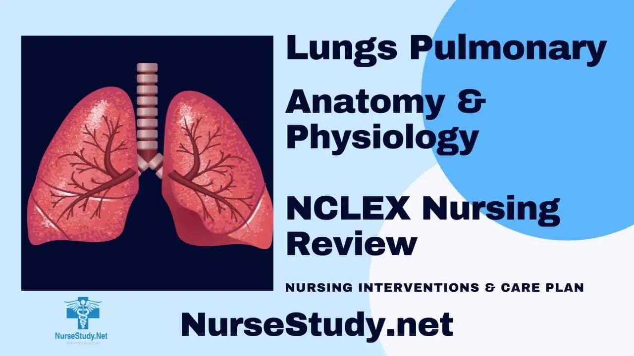
The lungs start from the root of the neck (apex) and lie on top of the diaphragm (base). This essential organ for respiration is protected by 12 ribs on each side and weighs around 1.3 kilograms. A double fold of a thin layer called pleura separates the lungs into lobes.
This pleura acts like a protective sheath, sort of like a thin membrane that surrounds the lungs and helps them expand when breathing in and out. The lungs have two lobes, with the right always being bigger and heavier than the left one as it needs to leave space for the heart. The right lobe also has three lobes, whereas the left only has two.
The right lobe consists of the superior lobe (located on the topmost), middle lobe, and inferior lobe; the left lobe consists of the superior and inferior lobe. Fissures separate each lobe in both pairs; the superior lobe and middle lobe are separated by a horizontal fissure, and the middle lobe and inferior lobe by an oblique fissure, similar to the left lung.
The blood supply in the lungs is supplied by two sources, the pulmonary and bronchial system of vessels, as the lungs are divided into supporting and functional parts. As the heart pumps out blood, the pulmonary circulation is responsible for transporting oxygen-poor blood to the lungs.
When it enters the lungs, the blood becomes oxygenated, which will then return to the heart. Differentiation of pulmonary circulation from systemic circulation is of paramount importance since instead of the mentioned mechanism, blood is supplied to the tissues in the body in the systemic circulation.
The process involves deoxygenated blood entering the right atrium, into the right ventricle, and then releasing carbon dioxide to be expelled (systemic) in exchange for oxygen in a process called respiration. Oxygen-rich blood departs from the lungs and enters through a series of pulmonary veins.
Basically, systemic circulation transports blood to the rest of the body, while pulmonary circulation is mainly grounded on two sites, the heart and the lungs.
Physiology of the Lungs in relation to the Respiratory System
To understand respiration, the significant path of entry and exit of air should be mentioned. It is essential to highlight that the nares, nasal cavities, meatuses, and other adjacent structures of the upper airway are lined with mucous membranes, hair follicles, and sebaceous glands.
Hence, when you breathe in air through the nose, your nasal cavity releases mucus that contains lysozymes; these are enzymes that kill bacteria. Interestingly, this process eliminates external antigens and debris from reaching the Respiratory zone, where the structures are solely dedicated to gas exchange.
The aforementioned process is one of many steps involved in respiration, specifically the filtering mechanism of the conducting zone and tracheobronchial tree. The first step during respiration, which is ventilation, occurs by taking up gas from the surrounding atmosphere to the balloon-like structures found in the lungs.
This is achieved via the contraction of accessory organs and structures involved in this process, but prior to its contraction, the CNS first sends signals to innervate the muscles involved in the respiratory airflow.
The diaphragm can be found below the thorax and it serves as the primary facilitator during breathing; it also functions by allowing muscle contraction and partial expansion of the lungs to happen in order to suction in air. To put it simply, as the diaphragm flattens and the chest expands, it creates a vacuum to draw in air.
This happens due to a small cavity just below the lungs. During inhalation (or inspiration), the lungs enclosed downward (filling in the cavity), causing an increased volume and decreasing pressure in the hollow space of the thorax—in contrast, with exhalation (or breathing out) where the muscles of the lungs and diaphragm relax.
Another probable reason why expiration is at a resting level is that the muscles of the upper airways are designed for inspiratory breathing.
The rib cage also plays a role during breathing since it also slightly expands with the help of several accessory respiratory muscles, the scalenes, and intercostal muscles.
During deep inhalation, the possible involvement of other accessory muscles can be observed, such as the parasternal intercostal muscles.
The lung’s central role in the body is gas exchange. During breathing, the body inhales oxygen that will enter the airways until it reaches the tiny sacs found in the lungs, called alveoli. Diffusion takes place due to partial pressure, and oxygen enters through the extracellular matrix by passing through the membranes between the alveoli and capillaries.
Partial pressure refers to a pressure gradient wherein gases flow from high pressure to low-pressure areas, and this mechanism permits air to flow in the lungs. Going back to passive diffusion of oxygen via external respiration (another term for gas exchange), oxygen diffuses into the deoxygenated blood.
On the other hand, carbon dioxide diffuses out of the deoxygenated blood, permitting the active exchange of both gases. One fact is that gas exchange happens at a rapid rate; simultaneously, capillary blood exchange fairs the same with its rate affected by the amount of cardiac output.
However, one thing to consider regarding ventilation-perfusion is the relatively small number of alveoli actually involved in gas exchange. And for that reason, the Ventilation/Perfusion ratio or the V/Q ratio serves as an additional measure for the efficiency of alveolar ventilation. This ratio varies all throughout the lungs; therefore, it can be variable. A mismatch V/Q ratio (mismatch ventilation and perfusion) is problematic since it translates to an existing problem in alveolar ventilation.
It can be a significant basis for the different types of lung failure and their probable cause. Hence, it is a key in determining ventilation efficacy and alveolar levels of carbon dioxide and oxygen. In healthy individuals, around .8 V/Q ratio is considered to be normal, but it may also depend on the region where the lung has been sampled.
Basically, matching perfusion and ventilation may indicate normal gas exchange. A below the normal (too low) perfusion where one lung is ventilated but the other is perfused denotes an alveolar dead space.
In contrast, a below the standard (too low) ventilation having a lower ratio denotes a shunt, indicating an inadequate supply of air or possibly an obstruction in the airway.
Diseases of the Lung
Despite effective mechanisms, the pulmonary and systemic circulation isn’t immune and invincible to foreign antigens or pathologic agents. There are many diseases of the lungs from Asthma, COPD, etc.
But one of the most severe problems affecting pulmonary circulation is a disease called pulmonary embolism. This disease is characterized by a blood clot that has been transported and has entered the lungs, causing an infarction. Infarction is characterized by tissue death, even from a section in the lung, caused by blockage of its blood supply.
Like what was mentioned, the blood clot would impede blood flow which may cause death or further complications to develop in the long run.
These blood clots have been determined to arise in the deep veins found in the legs (which is part of the systemic circulation) due to blood pooling from an injury, surgery, or even prolonged sitting. Since the leg veins are directed to the right portion of the heart, the clot is improbable to be broken down before it can reach the pulmonary circulation.
Upon reaching the pulmonary artery, the blood clot (or embolus) blocks the blood flow causing a decrease in blood pressure since the heart cannot pump enough blood. When this event happens, the heart may become strained, while the alveoli (responsible for gas exchange) may die due to the absence of blood supply.
The aftermath of an alveolar dead space and a decreased perfusion (meaning passage of blood) is expected. Signs and symptoms shortly follow after, presenting as difficulty in breathing or shortness of breath and episodes of chest pain.
This is a severe and life-threatening case, and if not treated immediately using fibrinolytics, as mentioned before, it can cause death, or the damage may become irreversible.
Nursing References
Ackley, B. J., Ladwig, G. B., Makic, M. B., Martinez-Kratz, M. R., & Zanotti, M. (2020). Nursing diagnoses handbook: An evidence-based guide to planning care. St. Louis, MO: Elsevier. Buy on Amazon
Gulanick, M., & Myers, J. L. (2017). Nursing care plans: Diagnoses, interventions, & outcomes. St. Louis, MO: Elsevier. Buy on Amazon
Ignatavicius, D. D., Workman, M. L., Rebar, C. R., & Heimgartner, N. M. (2018). Medical-surgical nursing: Concepts for interprofessional collaborative care. St. Louis, MO: Elsevier. Buy on Amazon
Silvestri, L. A. (2020). Saunders comprehensive review for the NCLEX-RN examination. St. Louis, MO: Elsevier. Buy on Amazon
Disclaimer:
Please follow your facilities guidelines, policies, and procedures.
The medical information on this site is provided as an information resource only and is not to be used or relied on for any diagnostic or treatment purposes.
This information is intended to be nursing education and should not be used as a substitute for professional diagnosis and treatment.