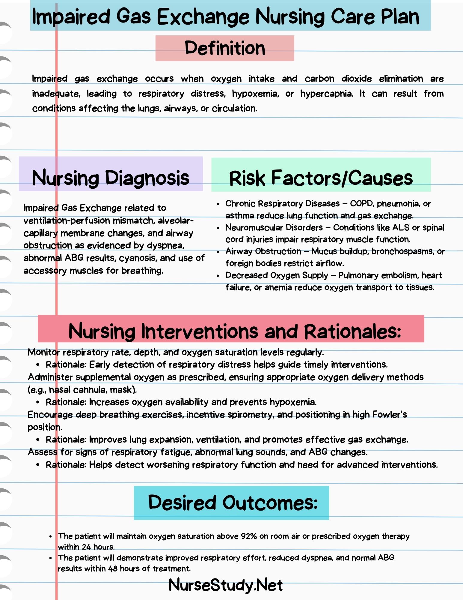
Impaired Gas Exchange is the NANDA-I nursing diagnosis for an excess or deficit in oxygenation and/or carbon dioxide elimination at the alveolar-capillary membrane. You see it as low oxygen (hypoxemia), high carbon dioxide (hypercapnia), or both: a falling SpO2, abnormal arterial blood gases, restlessness or confusion, fast breathing, and a fast heart rate. Common causes are COPD, pneumonia, heart failure with pulmonary edema, pulmonary embolism, and ARDS. Nursing care centers on upright positioning, oxygen titrated to the target the provider ordered, close monitoring of breathing and mental status, airway clearance, saving energy, and fast escalation when the client gets worse.
At a glance
- What it is
- A problem with moving oxygen into the blood and carbon dioxide out of it, at the level of the alveoli and the capillaries around them. The airways may be open and the client may be breathing hard, but the gases still do not cross well.
- Who gets it
- Clients with COPD or asthma flares, pneumonia, heart failure with fluid in the lungs, pulmonary embolism, ARDS, atelectasis after surgery, pulmonary fibrosis, and anyone whose breathing is slowed by opioids or sedatives.
- Key signs
- Early: restlessness, anxiety, fast breathing, fast heart rate, falling SpO2. With high CO2: headache, drowsiness, confusion. Late: cyanosis, slow heart rate, and a falling level of consciousness. The ABG confirms it.
- Nursing focus
- Sit the client up, give and titrate oxygen to the ordered target, watch SpO2, breathing effort, and mental status as trends, clear secretions, give ordered treatments for the cause, cluster care, and teach breathing techniques.
- Act now
- Call the rapid response team or provider for new confusion or drowsiness, SpO2 below the client’s target despite oxygen, inability to speak in full sentences, a tiring client, a very fast or very slow breathing rate, or cyanosis. Stay with the client.
Pathophysiology
Oxygen and carbon dioxide move between the alveoli and the pulmonary capillaries by diffusion across a very thin membrane. Gas exchange works when three things line up: air reaches the alveoli (ventilation), blood reaches the same alveoli (perfusion), and the membrane between them is thin and healthy. Impaired gas exchange happens when one or more of these fail.
- Ventilation-perfusion (V/Q) mismatch — the most common cause. Some alveoli get blood but little air (pneumonia, atelectasis, mucus plugging in COPD), or air but little blood (pulmonary embolism).
- Shunt — blood passes alveoli that get no air at all, such as fluid-filled or collapsed alveoli in ARDS or severe pneumonia. Hypoxemia from shunt improves only a little with extra oxygen.
- Diffusion problems — a thick or scarred membrane (pulmonary fibrosis), fluid in the interstitium and alveoli (pulmonary edema), or loss of surface area (emphysema).
- Hypoventilation — too little air moves in and out, so CO2 builds up and oxygen falls. Causes include opioid or sedative effects, a client too tired to keep breathing hard, and neuromuscular weakness.
Two patterns follow. In hypoxemic (type 1) respiratory failure, oxygen is low and CO2 is normal or low, as in pneumonia, pulmonary edema, or ARDS. In hypercapnic (type 2) failure, CO2 rises and causes respiratory acidosis, as in a COPD exacerbation or opioid-induced hypoventilation. Low oxygen first raises the heart and breathing rates to compensate; when the client tires or the brain is starved of oxygen, the level of consciousness falls and the heart rate can slow, which is a late and dangerous sign.
Choosing between the respiratory diagnoses
| Diagnosis (NANDA-I 2024–2026) | The problem is… | Typical clues |
|---|---|---|
| Impaired Gas Exchange | Oxygen and CO2 crossing the alveolar-capillary membrane | Abnormal ABG, low SpO2, restlessness, confusion, cyanosis |
| Ineffective Airway Clearance | Secretions or obstruction in the airways | Weak or ineffective cough, rhonchi, thick sputum |
| Ineffective Breathing Pattern | The rate, rhythm, or depth of breathing | Shallow, fast, or slow breathing; accessory muscle use; pursed-lip breathing |
| Impaired Spontaneous Ventilation | The client cannot keep breathing on their own | Rising CO2, falling SpO2, exhaustion, need for ventilatory support |
How it looks in common conditions
| Condition | Main mechanism | Nursing priority |
|---|---|---|
| COPD exacerbation | V/Q mismatch, air trapping, CO2 retention | Controlled oxygen to the ordered target, bronchodilators, watch for drowsiness from rising CO2 |
| Pneumonia | Exudate fills alveoli (V/Q mismatch, shunt) | Oxygen, airway clearance, antibiotics on time, watch for sepsis |
| Heart failure with pulmonary edema | Fluid in the interstitium and alveoli | Sit upright, oxygen, diuretics as ordered, daily weights, intake and output |
| Pulmonary embolism | Clot blocks blood flow (dead space) | Oxygen, anticoagulation as ordered, watch for low blood pressure and right heart strain |
| ARDS | Leaky, inflamed alveoli; severe shunt | ICU care, ventilator settings by protocol, prone positioning when ordered |
| Opioid or sedative effect | Hypoventilation | Sedation scale and respiratory rate checks, stop the drug and give the reversal agent per order or protocol |
Nursing assessment
Subjective data
- Shortness of breath at rest or on exertion; “I can’t get enough air”
- Needing to sit up or sleep in a chair to breathe (orthopnea)
- Headache on waking, sleepiness, or trouble thinking (possible CO2 retention)
- Fatigue, weakness, and anxiety or a feeling that something is very wrong
- Cough, sputum amount and color, chest pain, fever
- History of COPD, asthma, heart failure, clotting, recent surgery or immobility, and smoking
- Home oxygen use and the client’s usual SpO2 and activity level
- Recent opioids, sedatives, or sleep medicines
Objective data
- Respiratory rate, depth, rhythm, and effort; accessory muscle use, nasal flaring, retractions, tripod position
- Ability to speak: full sentences, phrases, or single words
- SpO2 trends at rest and with activity, with the oxygen device and flow rate recorded
- Breath sounds: crackles, wheezes, rhonchi, or diminished or absent sounds
- Heart rate and rhythm, blood pressure, temperature
- Level of consciousness and orientation, restlessness, and a sedation score if opioids are given
- Skin and mucous membranes: pallor, diaphoresis, cyanosis of the lips and tongue
- Arterial blood gas results, hemoglobin, chest X-ray, and other tests the provider orders
Reading the ABG
| Value | Typical adult range | In impaired gas exchange |
|---|---|---|
| pH | 7.35–7.45 | Below 7.35 with high CO2 = respiratory acidosis |
| PaCO2 | 35–45 mmHg | High in hypoventilation and COPD; may be low early on from fast breathing |
| PaO2 | 80–100 mmHg on room air | Low (hypoxemia) |
| HCO3− | 22–26 mEq/L | Above range when the kidneys have compensated for long-standing CO2 retention |
Reference ranges vary slightly between laboratories, and older adults and clients with chronic lung disease may have a different baseline. Always compare results with the client’s own baseline and the clinical picture.
Nursing diagnoses used with impaired gas exchange
- Impaired Gas Exchange — abnormal oxygen and/or CO2 levels from V/Q mismatch or membrane changes
- Ineffective Airway Clearance — secretions the client cannot clear
- Decreased Activity Tolerance — oxygen supply cannot meet demand during activity
- Anxiety — fear and distress from breathlessness
- Ineffective Breathing Pattern — fast, shallow, or labored breathing
- Impaired Spontaneous Ventilation — the client cannot keep breathing without support
- Fatigue — exhaustion from the work of breathing and poor sleep
- Inadequate Health Knowledge — gaps in knowing about inhalers, oxygen, or warning signs
Worked care plans
Four plans in the same ADPIE shape. The scenario is a 71-year-old woman with COPD admitted with an exacerbation and right lower lobe pneumonia. She is sitting forward with her hands on her knees, using her neck muscles to breathe, and can speak only three or four words at a time. Temperature 38.4 °C (101.1 °F), heart rate 112, respiratory rate 28, blood pressure 148/86, and SpO2 86% on room air. She has rhonchi and crackles in the right lower lobe and expiratory wheezes throughout. Her ABG on room air shows pH 7.33, PaCO2 54 mmHg, PaO2 52 mmHg, and HCO3− 28 mEq/L. The provider orders oxygen by nasal cannula titrated to an SpO2 of 88–92% for her, nebulized bronchodilators, a corticosteroid, antibiotics, and a repeat ABG. She says, “I can’t catch my breath. Please don’t leave me.”
1. Impaired Gas Exchange
Problem-focusedSubjective: “I can’t catch my breath.” “My head is pounding.” “I’m so tired.”
Objective: SpO2 86% on room air; ABG pH 7.33, PaCO2 54 mmHg, PaO2 52 mmHg; respiratory rate 28; heart rate 112; restless and picking at the bed sheets; dusky lips; accessory muscle use.
Impaired Gas Exchange as evidenced by hypoxemia, hypercapnia, abnormal arterial pH, restlessness, tachycardia, abnormal skin color, and headache; associated conditions: ventilation-perfusion imbalance and alveolar-capillary membrane changes (COPD exacerbation with right lower lobe pneumonia).
- SpO2 stays within the target range the provider ordered for her
- Repeat ABG shows improving pH and CO2 compared with admission
- Client is alert, oriented, and less restless, with a slower breathing rate and less accessory muscle use
- Raise the head of the bed and let her lean forward on an over-bed table if that is easier
- Start oxygen as ordered and titrate to her prescribed target range; do not withhold oxygen from a hypoxemic client
- Monitor SpO2 continuously, and check respiratory rate, effort, breath sounds, and level of consciousness often, recording the oxygen device and flow each time
- Give the ordered bronchodilators, corticosteroid, and antibiotics on time
- Draw or arrange the repeat ABG as ordered and compare it with the first
- Report rising drowsiness, a falling pH or rising CO2, or SpO2 below target to the provider; respiratory therapy and the provider decide on noninvasive ventilation
- Hold sedatives and check with the provider before giving any opioid or sleep medicine
After 24 hours on 2 L/min by nasal cannula, SpO2 was 90%, respiratory rate 22, and heart rate 96. The repeat ABG showed pH 7.38 and PaCO2 49 mmHg. She was alert, oriented, and speaking in full sentences, and her lips were pink. Goal met; continue monitoring.
2. Ineffective Airway Clearance
Problem-focusedSubjective: “It’s stuck in my chest and I can’t get it up.” “Coughing wears me out.”
Objective: Weak, ineffective cough; thick yellow sputum; rhonchi in the right lower lobe that change after coughing; expiratory wheezes; respiratory rate 28.
Ineffective Airway Clearance related to retained secretions and excessive mucus as evidenced by ineffective cough, adventitious breath sounds, excessive sputum, dyspnea, and altered respiratory rate; associated conditions: COPD and respiratory infection.
- Client coughs up secretions effectively
- Rhonchi clear or decrease after coughing
- Client shows the huff cough correctly
- Keep her upright, and time coughing and airway clearance after her bronchodilator treatment
- Teach the huff cough: a medium breath in, then two or three forceful “huffs” with an open mouth
- Encourage oral fluids unless she has a fluid limit, to keep secretions thinner
- Work with respiratory therapy on airway clearance devices, such as an oscillating positive expiratory pressure device, if ordered
- Collect a sputum specimen as ordered, ideally before the first antibiotic dose
- Suction only if she cannot clear secretions herself, following facility policy
- Note sputum amount, color, and thickness each shift
By day 2 she huffed and coughed up moderate amounts of thinner sputum after each treatment, and the rhonchi cleared after coughing. Wheezes decreased. Goal met.
Free download
NCLEX Readiness Diagnostic
Fifty questions with a written rationale on every option, scored by section so you can see which content area is weakest. Sent as a PDF.
3. Decreased Activity Tolerance
Problem-focusedSubjective: “Just walking to the bathroom wipes me out.” “I have to stop and rest.”
Objective: On day 2, SpO2 fell from 90% to 84% and heart rate rose from 96 to 128 while walking about 6 meters (20 feet) to the bathroom; she stopped twice and needed 5 minutes to recover.
Decreased Activity Tolerance related to imbalance between oxygen supply and demand and physical deconditioning as evidenced by exertional dyspnea, abnormal heart rate response to activity, and fatigue.
- Client walks to the bathroom with SpO2 staying within her target range and heart rate returning to her resting level soon after
- Client uses pursed-lip breathing and planned rests during activity
- Client names three ways to save energy at home
- Cluster care and plan rest periods before and after activity and meals
- Check SpO2, heart rate, and breathlessness before, during, and after activity, and stop if she meets the stopping criteria in the facility or PT protocol
- Ask the provider whether her oxygen order covers a higher flow during activity
- Offer a bedside commode until she can walk to the bathroom safely
- Coach pursed-lip breathing and exhaling during effort, such as when standing up
- Increase activity a little each day and request a physical therapy evaluation
- Teach energy conservation: sit to wash and dress, keep things within reach, and rest between tasks
By day 4 she walked to the bathroom and back with one rest. SpO2 stayed at 89–91% on her ordered oxygen, and her heart rate returned to resting within 3 minutes. She described sitting to shower and spreading chores across the day. Goal met; PT continues.
4. Anxiety
Problem-focusedSubjective: “Please don’t leave me.” “I feel like I’m going to suffocate.” “What if this is it?”
Objective: Restless, gripping the bed rails, pulling at the nasal cannula, wide-eyed, and asking the same questions again; breathing faster when frightened.
Anxiety related to stressors (acute breathlessness and an unfamiliar hospital setting) as evidenced by expressed fear, restlessness, and increased respiratory rate.
Restlessness can be a sign of hypoxia, not only anxiety. Check SpO2, breathing, and mental status first, every time.
- Client states she feels calmer and safer
- Client uses pursed-lip breathing when she feels breathless
- Client keeps her oxygen on
- Rule out worsening hypoxia or rising CO2 as the cause of restlessness before treating it as anxiety
- Stay with her during episodes of breathlessness, speak slowly and calmly, and use short sentences
- Coach pursed-lip breathing with her, breath by breath
- Offer a handheld fan directed at her face, which can ease the feeling of breathlessness
- Explain each treatment and why it helps, and keep the call light within reach
- Avoid giving sedatives for anxiety unless the provider has evaluated her, because they can slow breathing and raise CO2
Her oxygen level was within target when she was restless, so the cause was fear. With coaching she slowed her breathing, kept the cannula on, and said, “It helps when you breathe with me.” Later she started pursed-lip breathing on her own. Goal met.
Interventions and rationales
Client and family teaching
- Pursed-lip breathing: breathe in through your nose, then breathe out slowly through pursed lips, taking about twice as long to breathe out as to breathe in
- Use your inhalers the way you were shown, and bring them to every appointment so the technique can be checked
- Follow your written action plan: know your usual symptoms, your warning signs, and who to call for each
- If you use home oxygen, use the flow your provider prescribed; never smoke or let anyone smoke near it, and keep it away from open flames and heat
- Stop smoking and avoid secondhand smoke; ask about medicines and programs that help you quit
- Ask your provider which vaccines are recommended for you, such as flu and pneumonia vaccines
- Pace yourself, rest between tasks, and do the exercises from pulmonary rehabilitation if it was recommended
- Call 911 for severe shortness of breath, trouble speaking, blue lips or fingertips, chest pain, or confusion or sleepiness that is new
NurseStudyPrep.com
You’ve read the care plan. Now answer one.
A priority item in the NCLEX format. On Prep, every option gets a written rationale — including the three that are wrong.
A client with a COPD exacerbation is receiving oxygen by nasal cannula, titrated to the SpO2 target the provider ordered. The flow was increased 2 hours ago. Which finding should the nurse act on first?
Why C. New drowsiness in a client with COPD can mean CO2 is rising (hypercapnia), which can lead to respiratory failure. The nurse stays with the client, assesses breathing and airway, and calls the provider or rapid response team; an ABG is likely. The nurse does not simply turn the oxygen off. Option A is the expected result. Option B is a helpful technique the client uses to ease breathing. Option D is a long-standing finding in COPD. Prep also explains why each wrong option looks reasonable.
7 days for $1, then $19 a month, $49 for three months or $129 for the school year. Cancel any time.
Frequently asked questions
What is impaired gas exchange?
It is a NANDA-I nursing diagnosis for an excess or deficit in oxygenation and/or carbon dioxide elimination at the alveolar-capillary membrane. In practice it means the blood is not picking up enough oxygen, not getting rid of enough CO2, or both.
What is the difference between impaired gas exchange and ineffective airway clearance?
Ineffective Airway Clearance is a problem in the airways, such as secretions the client cannot cough up. Impaired Gas Exchange is a problem at the alveoli, where gases cross into and out of the blood. One can lead to the other: retained secretions can cause impaired gas exchange.
What are the early signs of impaired gas exchange?
Restlessness, anxiety, fast breathing, a fast heart rate, and a falling SpO2. Cyanosis, a slow heart rate, and a falling level of consciousness are late signs.
What is the priority nursing intervention for impaired gas exchange?
Assess airway and breathing, position the client upright, and give oxygen as ordered to reach the client’s target SpO2, then monitor closely and report deterioration. Treating the cause follows.
Should oxygen be limited in a client with COPD?
Oxygen is titrated, not withheld. For clients at risk of CO2 retention, providers often set a lower SpO2 target, and the nurse adjusts oxygen to stay in that range and watches for drowsiness or a rising CO2. A hypoxemic client always receives oxygen.
How do you write an impaired gas exchange nursing diagnosis?
State the label, the evidence, and the associated conditions, for example: “Impaired Gas Exchange as evidenced by hypoxemia, hypercapnia, and restlessness; associated condition: ventilation-perfusion imbalance.” Some programs use “related to” for the cause instead.
References
- Herdman, T. H., Kamitsuru, S., & Lopes, C. T. (Eds.). NANDA International Nursing Diagnoses: Definitions and Classification, 2024–2026. Thieme.
- Global Initiative for Chronic Obstructive Lung Disease. Global Strategy for the Diagnosis, Management, and Prevention of COPD. GOLD report, current edition.
- O’Driscoll, B. R., Howard, L. S., Earis, J., & Mak, V. BTS guideline for oxygen use in adults in healthcare and emergency settings. Thorax, 2017;72(Suppl 1):ii1–ii90.
- U.S. Food and Drug Administration. Pulse Oximeter Accuracy and Limitations: FDA Safety Communication. 2021.
- Ackley, B. J., Ladwig, G. B., Makic, M. B. F., Martinez-Kratz, M. R., & Zanotti, M. Nursing Diagnosis Handbook: An Evidence-Based Guide to Planning Care (13th ed.). Elsevier, 2023.
- Harding, M. M., Kwong, J., Hagler, D., & Reinisch, C. Lewis’s Medical-Surgical Nursing: Assessment and Management of Clinical Problems. Elsevier.
- Hinkle, J. L., Cheever, K. H., & Overbaugh, K. J. Brunner & Suddarth’s Textbook of Medical-Surgical Nursing. Wolters Kluwer.
- Silvestri, L. A. Saunders Comprehensive Review for the NCLEX-RN Examination. Elsevier, 2023.
SpO2 targets, oxygen devices and flow rates, ABG reference ranges, activity stopping criteria, sedation scales, and medicine choices vary by client, by provider, and by facility. Follow the provider’s orders, the client’s individual targets, and your own institution’s policies and protocols. This page supports study and does not replace course instruction or clinical judgment.
